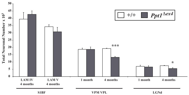
Figure 8.
Early loss of thalamic relay neurons in Ppt1Δex4 knock-in mice. Histograms of unbiased optical fractionator estimates of the number of Nissl stained lamina IV granule neurons and lamina V projection neurons in somatosensory barrelfield (S1BF) cortex; the ventral posterior nucleus of the thalamus (VPM/VPL), which provides afferent input to S1BF; and visual relay neurons in the dorsal lateral geniculate thalamic nucleus (LGNd) of Ppt1Δex4 mice and age-matched controls (+/+) at different stages of disease progression. No significant loss of cortical neurons in laminae IV or V of S1BF was evident in Ppt1Δex4 mice at either 1 or 4 months of age. In marked contrast, and consistent with the phenotype of Ppt1-/- mice (Kielar et al., 2007), a significant loss of both VPM/VPL and LGNd neurons was already observed at 4 months of age. (* p<0.05; *** p<0.001, ANOVA with post-hoc Bonferroni analysis).

