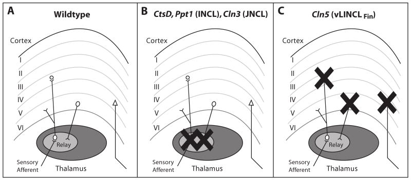
Figure 9.
Contrasting patterns of corticothalamic neuron loss in NCL mouse models. (A) Normal organization of thalamocortical pathways. Ascending sensory afferents terminate upon thalamic relay neurons, with one relay nucleus for information of different modalities. These relay neurons project mainly to lamina IV granule neurons of the appropriate cortical region, with collateral innervation of lamina VI. In turn, the cortex provides feedback projections to the thalamus from lamina VI, and lamina V neurons project outside this cortical region. (B, C) The thalamocortical system of all NCL mouse models displays progressive neuron loss. However, the timing and sequence in which these pathways are affected differs radically between forms of NCL. (B) In Cathepsin D, Ppt1 and Cln3 deficient mouse models the neuron loss begins in the thalamus and only subsequently occurs in the corresponding cortical region. (C) In contrast, neuron loss progresses in a completely opposite sequence in Cln5 deficient mice. As this study reveals, neuron loss is first evident in the cortex of Cln5-/- mice, with loss of thalamic relay neurons occurring later in disease progression. However, regardless of the pattern of cell loss, visual pathways are consistently affected first in all NCL mouse models.

