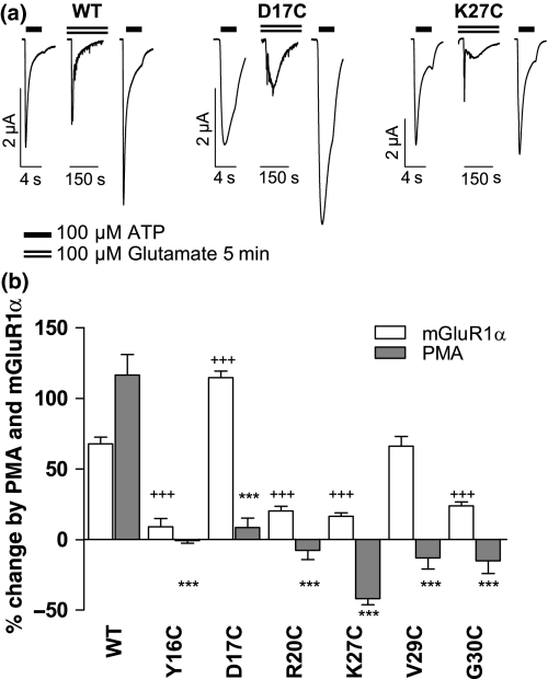
Fig. 5.
The effects of mGluR1α receptor activation on P2X1 receptor mutants. (a) Sample traces for a given oocyte co-expressing either WT P2X1, D17C or K27C mutant P2X1 receptor with mGluR1α receptors. Responses to a maximal concentration of ATP (100 μM, indicated by bar) are shown before and after the application of glutamate (dotted line). Glutamate (100 μM) evoked an inward calcium activated chloride current and potentiated subsequent ATP evoked responses for WT and D17C mutants but not for the K27C mutant P2X1 receptor. (b) The effects of mGluR1α receptor (100 μM glutamate) and PMA (100 nM) on WT and the cysteine mutants are shown (+++p< 0.001 comparing mGluR1 receptor regulation of mutants to WT, ***p< 0.001 for mutants treated with PMA compared to the WT effect). For most of the mutants unable to exhibit the PMA potentiation, no potentiation was seen following the activation of mGluR1α receptor. However, the mGluR1α receptor stimulated potentiation was still robust for the D17C and V29C mutants.

