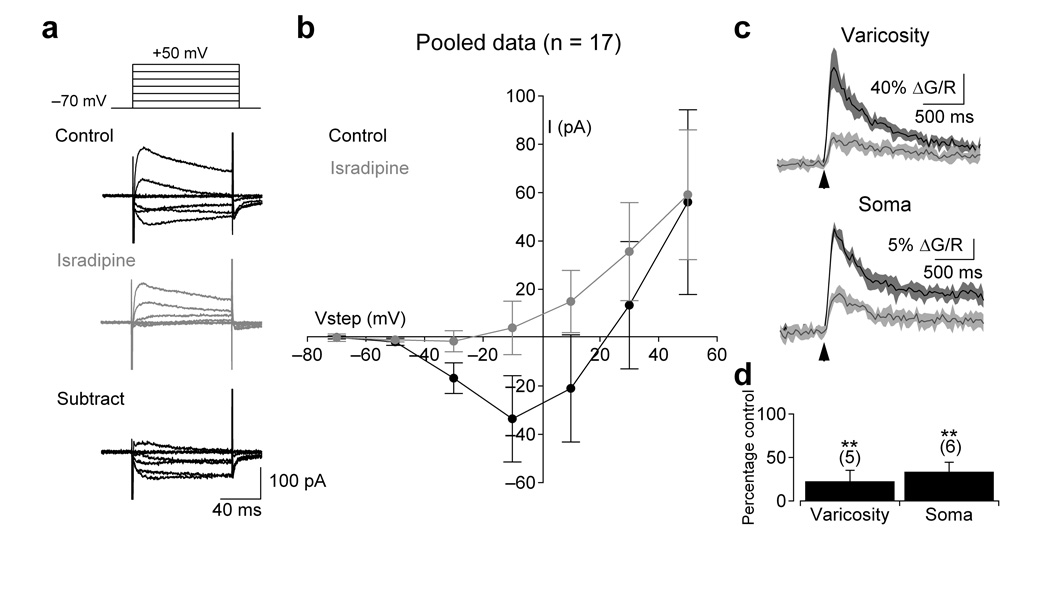
Figure 2.
Functional L-type VGCCs are expressed at A17 synaptic varicosities and somata. (a) A series of depolarizing voltage steps (−70 to +50 mV in 20 mV increments; 100 ms) elicited an isradipine (10 µM)- sensitive inward current. (b) Summary of the current-voltage relationship in control and in the presence of isradipine (10 µM; n = 17 cells). (c) Depolarizing voltage steps (100 ms to −10 mV) also elicited isradipine-sensitive fluorescence transients in the varicosities (top) and somas (bottom) of A17 amacrine cells. Fluorescence from a single varicosity (typically a 16×16 frame) was acquired at ~50 Hz and a small region of interest (as in Fig. 1b) was drawn around the varicosity to produce an average pixel value. Traces are the average of eight responses to 100 ms voltage steps before and after isradipine application (arrows indicate onset of step). Shaded regions indicate ± SD. (d) Summary of the effects of isradipine on voltage-dependent fluorescence at individual varicosities (n = 5 cells) and somas (n = 6 cells). Data in panels b and d represent mean ± SD.

