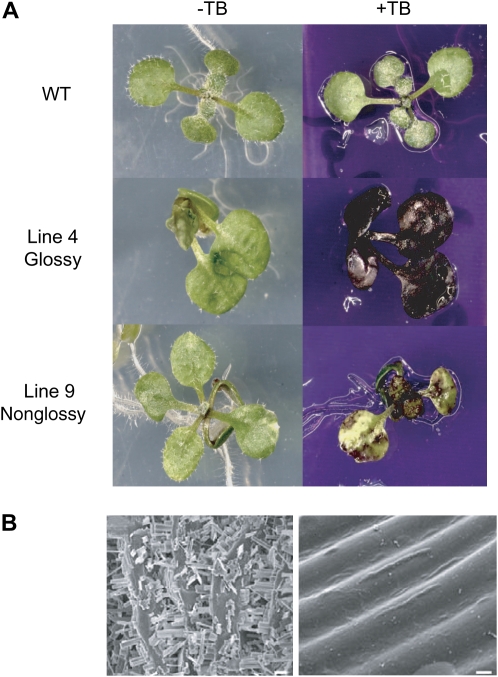
Figure 10.
AtKCR1-RNAi plants display cuticular defects. A, Two-week-old wild-type (WT) and AtKCR1-RNAi seedlings stained with TB. B, Epicuticular wax crystal structure on stem surfaces visualized by scanning electronic microscopy. Shown are images of the wild type (left panel, bar =10 μm) and glossy AtKCR1-RNAi line 4 (right panel, bar = 2 μm).

