Figure 7.
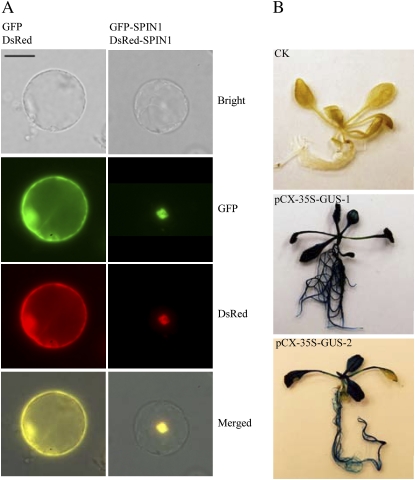
Protein subcellular localization and promoter activity analysis using the ZeBaTA vectors. A, Fluorescence microscopy of the coexpression of GFP and DsRed, or GFP-SPIN1 and DsRed-SPIN1 fusions in rice protoplasts. Scale bar = 20 μm. The RNA binding nuclear protein SPIN1 was used as a tester (Vega-Sánchez et al., 2008). B, GUS staining of Arabidopsis transformed with pCX-35S-GUS, where the 35S promoter was cloned into the vector pCXGUS-P to test the system. CK, Plant transformed with control vector pCAMBIA1300 (www.cambia.org); pCX-35S-GUS-1 and pCX-35S-GUS-2, two independent primary transgenic plants.

