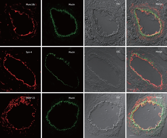
Figure 3.
Apical SNARE localization in unstimulated Brunner’s gland acini. Confocal images of unstimulated Brunner’s gland acini labeled with anti-mucin antibody (in green) and double labeled with anti-Munc18c, Syntaxin-4 or SNAP-23 antibodies (in red), and an overlay (merge) of the two with the DIC, which is also separately shown. Note the apical plasma membrane staining of Munc18c (and SNAP-23 and Syntaxin-4) and mucin accumulation beneath it. Scale bars correspond to 10 μm.

