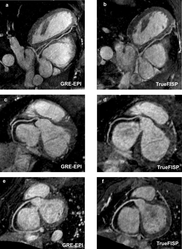
Figure 5.
Reformatted coronary artery images from 2 volunteers using the contrast-enhanced GRE-EPI acquisition (5a, c and e) and the ideal TrueFISP acquisition with an acceleration factor of 2, leading to longer imaging time (5b, d and f). Both the sequences show similar depiction of the coronary arteries, however, the imaging time for the contrast-enhanced GRE-EPI acquisition is reduced by a factor of 2 compared with the TrueFISP acquisition.

