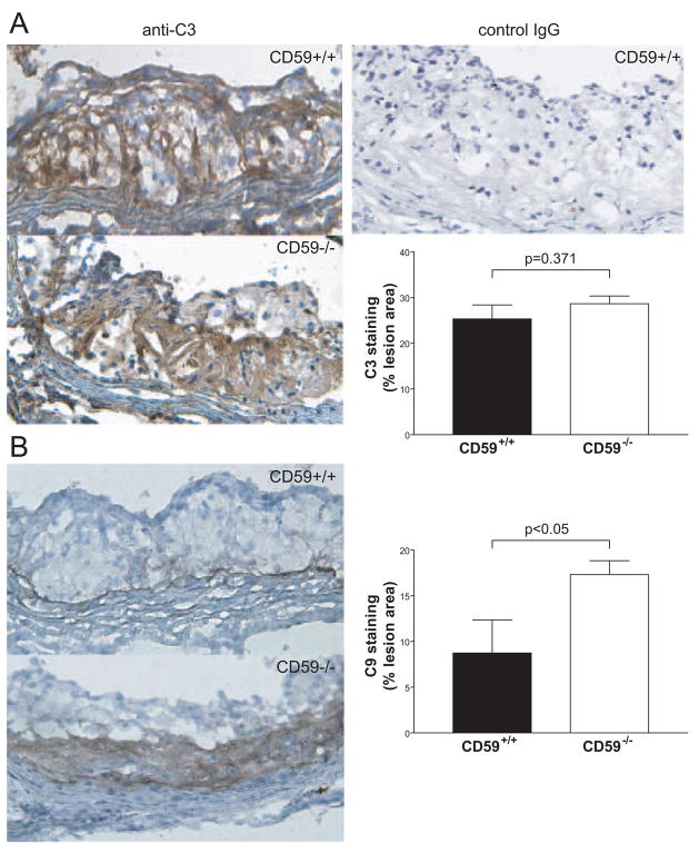
Figure 5.
CD59 deficiency increased the deposition of membrane attack complex within the atherosclerotic lesions. A, C3 staining of aortic root sections from female CD59−/− ApoE−/− and CD59+/+ ApoE−/− mice after 8 weeks of high fat diet (n=5 in each group). Left, representative photographs of sections stained for C3. Upper right, background staining with a goat IgG isotype control antibody. Lower right, morphometric analysis of C3-stained area in the lesions. B, C9 staining of aortic root sections from female CD59−/− ApoE−/− and control mice after 8 weeks of high fat diet (n=7 in each group). Left, representative photographs of sections stained for C9. Right, quantitative analysis of the C9 stained area of lesions. Results are expressed as the percentage of positive area. Lesion areas in 6 sections for each mouse were measured. Bars represent mean±SEM. P values are by Student t test.

