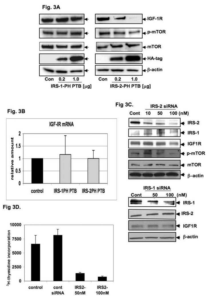
Fig. 3. IRS-2, but not IRS-1, in IGF-1R overexpression in PCA.
Total cell lysates from AsPC-1 cells transiently transfected with 0.2 and 1.0 µg of expression vector containing either (A) IRS-1 PH-PTB or IRS-2 PH-PTB or empty vector (control) were analyzed by immunoblotting with anti-IGF-1R 2C8 (IGF-IR) and anti-phospho-mTOR (phos-mTOR). Anti-HA tag (HA-tag) was measured to monitor transfection efficiency, β-actin served as control protein. (B) IGF-1R mRNA expression was analyzed from total RNA extracted from AsPC-1 cells transiently transfected with 1.0 µg of expression vectors containing either IRS-1 PH-PTB or IRS-2 PH-PTB and empty vector (control) were analyzed by RT-PCR using beta-actin as control. (C) siRNA of IRS-2, but not IRS-1 inhibits IGF-1R protein expression. Total cell lysates from AsPC-1 cells transiently transfected with siRNA of IRS-2; siRNA of IRS-1 or control siRNA (control) (all purchased from Santa Cruz) analyzed by immunoblotting with anti-IGF-1R 2C8 (IGF-1R) EGFR and other proteins. siRNA of IRS proteins did not influence each other. β-actin served as control protein. (D) 3H-thymidine incorporation of AsPC-1 cells after treating with IRS-2 siRNA (50nM and 100 nM) which showed that significant inhibition of cell proliferation.

