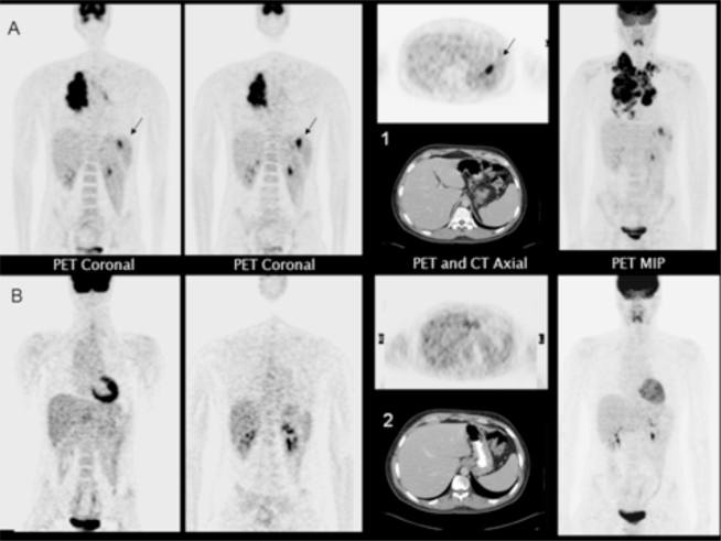
Figure 1 -.
A 22-year old male patient with conventional clinical stage II disease. A) FDG-PET coronal, axial and maximum intensity projection (MIP) images showed additional splenic lesions (arrows) not observed in the CT axial image (1). B) After two ABVD cycles, all lesions, including the splenic foci, disappeared; normal CT axial image (2).

