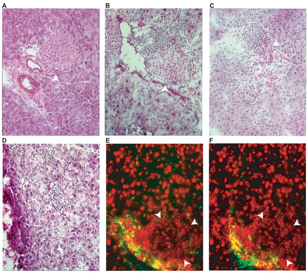
Figure 1. Pancreas histology of protein-immunized and DNA-immunized mice.

Frozen sections of pancreas were stained with hematoxylin and eosin (A-D) or immune-stained for CD4 (E) or CD8 (F). (A), DNA-immunized non-transgenic mouse pancreas, (B), protein-immunized DQ8-RIP7-hGAD65, double-transgenic mouse pancreas. (C-D), DNA-immunized DQ8-RIP7-GAD65 double-transgenic mouse pancreas. Note the more aggressive characteristic of the lymphocytic infiltration in C (some islet tissue preserved), and D (complete islet destruction) of the GAD65 DNA-immunized double-transgenic as compared with the peri-insular infiltration of protein-immunized double-transgenic mouse (B). Confluent nuclei in E and F (red) correspond to islet area (arrow delineated). Islets are indicated with arrowheads.
