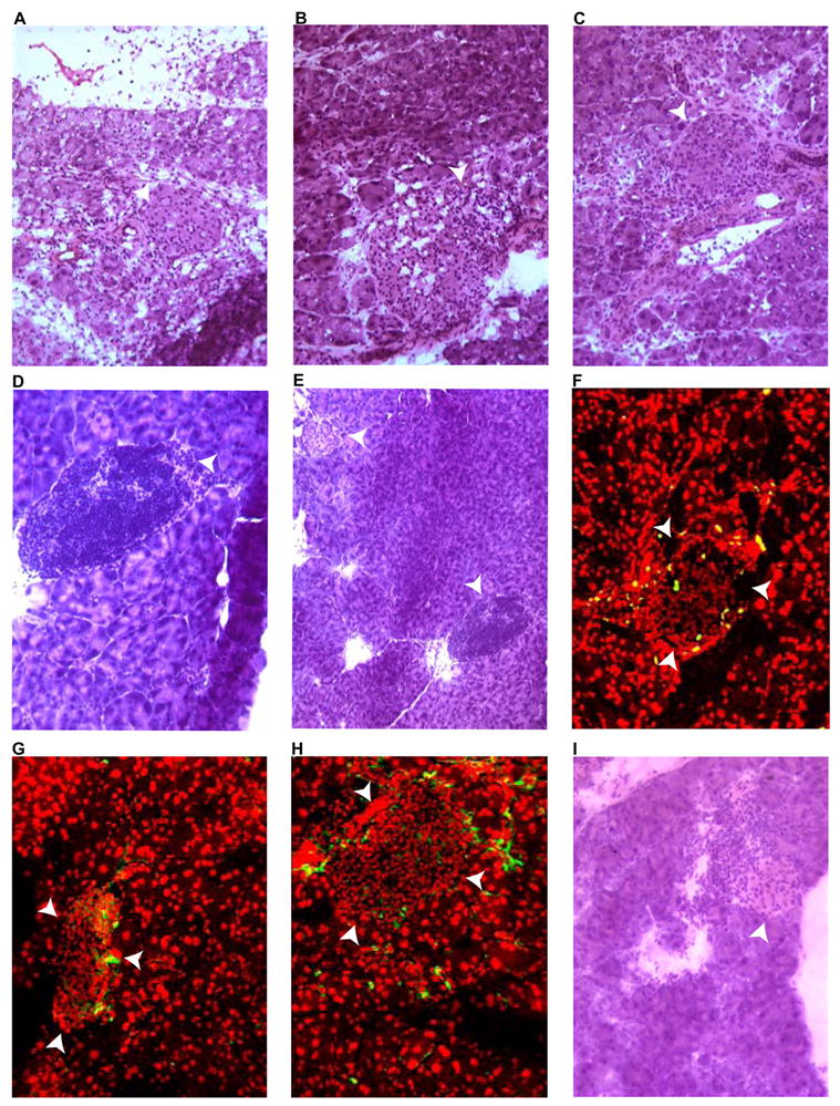
Figure 5. Progression of insulitis in DNA-immunized double-transgenic mice.

Frozen pancreatic sections, obtained at different time points following DNA immunization, were stained with hematoxilin and eosin (A-E and I), anti-BrdU (F), anti-CD4 (G), or anti-CD8 (H). White arrowheads indicate islets. A, eleven days post-immunization (infiltration present). B, week 5 post-immunization pancreas appeared massively infiltrated. C, week 9 post-immunization (more localized infiltration noted). D, week 13 post-immunization specimen shows a severely infiltrated islet. E: panoramic view of D showing the concomitant presence of normal islet (top left corner). F, post-BrdU treatment of an animal euthanized at week 17 (proliferating cells are exclusively detected in the islet). G and H, pancreas 25 weeks post-immunization (G=anti-CD4, H=anti-CD8). I, pancreas section of a recipient of adoptively transferred splenocytes 5 weeks post transfer.
