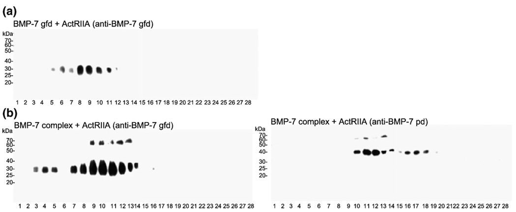
Fig. 6.
ActRIIA also displaces the BMP-7 pd. (a) Sucrose gradient of ActRIIA together with the BMP-7 gfd. Immunoblotting with anti-BMP-7 gfd showed a main peak in fractions 8 and 9, indicating the formation of a receptor-gfd complex, (b) Interaction of BMP-7 complex with ActRIIA. Immunoblotting of sucrose gradient fractions revealed the appearance of four peaks: free complex (fractions 11–14); complex bound to the receptor molecule (fractions 10–12); free gfd bound to the receptor (fractions 7–9); and high-molecular-weight complexes, caused by dimerization of the Fc domains of the receptor molecules, possibly consisting of two BMPRII-Fc dimers and multiple BMP-7 gfds (fractions 3–5).

