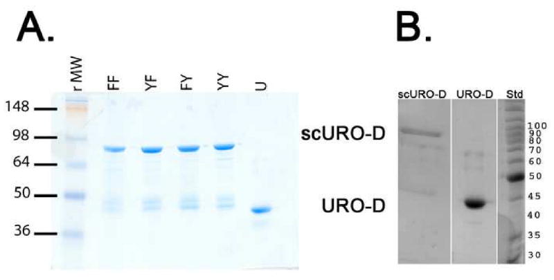
Figure 2. Purified proteins.

(A)1 μg sample of each of the purified proteins visualized on Coomassie-stained SDS-PAGE: scURO-D (YY), scURO-D(YF) (YF), scURO-D(FY) (FY), scURO-D(FF) (FF), and URO-D (U). The scURO-D proteins have an apparent molecular weight of 84 kDa while native URO-D runs at 42 kDa. Densitometry of the bands present in the scURO-D sample indicated that approximately 10 to 20% of the URO-D protein is present at the molecular weight corresponding to monomeric URO-D. (B) Crystals of scURO-D were removed from the crystallization well, washed and dissolved in 50 mM Tris pH 7.5. Proteins were separated by SDS page. Densitometry of the bands present in the scURO-D sample indicated that approximately 22% of the protein is present at a molecular weight corresponding to monomeric URO-D.
