
Fig. 1. Golgi complex is an additional MTOC.
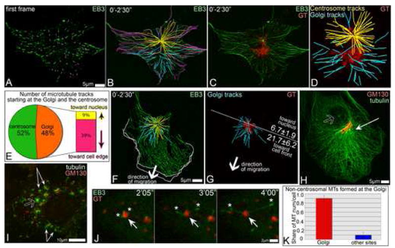
A-D. Detection of Golgi-originated MTs in time lapse recording of GFP-EB3 and mCherry-GT expressing RPE1 cell (5sec/frame). A. GFP-EB3 in the first frame of the video (green). Currently growing MTs are marked by magenta dots. B. Overlaid GFP-EB3 showing MT tracks within 2.5′. Magenta, tracks started at the frame one. Yellow, centrosomal tracks. Cyan, non-centrosomal tracks. C. Overlaid GFP-EB3 (green) and mCherry-GT (red) images within 2.5′. D. centrosomal (yellow) and non-centrosomal (cyan) MT tracks in the cell center and their relation to the Golgi position (GT, red). E. Percentage and directionality of Golgi-associated tracks (583 tracks in 10 cells, analyzed as above). F, G. Directionality of Golgi-associated tracks in a motile cell. Arrows indicate the direction of the cell re-location. F. Overlaid mCherry-EB3 tracks (2.5′, false-colored green). Centrosomal (yellow) and Golgi-associated (cyan) tracks in the cell center are shown. Outlines of the protruding cell front in the first and the last frame of the 5′ video are shown as white lines. G. Golgi (YFP-GT, false-colored red) and associated tracks (cyan). A line is drawn perpendicular to the direction of cell movement. Average number of MTs in 9 cells growing forwards or backwards are shown. H. Prominent MT array (thin arrow) in a polarized cell is rather associated with the Golgi (red) than with the centrosome (hollow arrow). Tubulin (green) and GM130 (red), immunostained. I, RPE1 cell fixed and stained 45″ after nocodazole washout. MTs (green) radiate from the Golgi mini-stacks (thin arrows) and the centrosome (hollow arrow). Tubulin, green. GM130, red. J. Live cell images of nocodazole washout. GFP-EB3-rich plus tips (green, asterisks) grow away from the mCherry-GT-marked Golgi stack (arrow, red). K. Number of non-centrosomal MTs nucleated at the Golgi (red) and elsewhere (blue) after nocodazole washout per cell, based on live recordings of mCherryEB3 and YFP-GT expressing cells (521 MTs in 8 cells).