
Fig. 3. CLASPs specifically localize to TGN membranes via GCC185 binding.
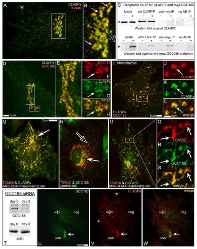
A,B, CLASPs (green) co-localize the TGN46 (red, thin arrows in B) along with the MT tips (star in A) and the centrosome (hollow arrow in B). Box in A is enlarged in B. C, Co-IP of CLASP2 and myc-GCC185 using either anti-CLASP2 or anti-myc antibodies. Upper panel, CLASP2. Lower panel, myc-GCC185. Non-transfected cells are marked as “–”, myc-GCC185-transfected cells as “+”. CLASP2 is co-precipitated from transfected cells by anti-myc antibody (Anti-myc IP). Myc-GCC185 is co-precipitated from transfected cells by anti-CLASP antibody (Anti-CLASP IP). Antibody-free beads used as control (No AB IP). D-H, CLASP co-localizes with ectopically expressed myc-tagged GCC185 at the TGN. Myc (red), CLASP (green). E, Enlarged tall box from D. F-H, Enlarged small box from D. F, myc-GCC185. G, CLASP. H, merge. Arrows, reference point. I-L, association of CLASPs and GCC185 is preserved in nocodazole. J-L, Enlarged small box from I. J, myc-GCC185. K, CLASPs. L, merge. Arrow, reference point. M, CLASP2-dTOM20 chimera (mito-CLASP) and mCherry-dTOM20 (red) co-expressing cell. CLASP staining (green) reveals both mito-CLASP (arrow, co-localized with mCherry-dTOM20 at mitochondria) and endogenous CLASPs. N. GCC185 at the Golgi (green, white arrow) does not co-localize with mCherry-dTOM20 at mitochondria (red, hollow arrow) in control cells. O-S. Mito-CLASP expressing cell. Endogenous GCC185 (green) is recruited to mitochondria (mCherry-dTOM20, red). Box is enarged in Q-S. Q, mCherry-dTOM20. R, GCC185. S, merge. Arrows, reference points. T, GCC185 is 90% depleted from RPE1 cells by siRNA. U-W, Mixed culture of GCC185-depleted (neg) and control (pos) cells. U, GCC185. V, CLASPs. W, merge of U and V. CLASP is localized to the Golgi in control cell (white arrow) but not in GCC185-depleted cell (hollow arrow). MT tip colalization of CLASP is intact in both cells (asterrisks). All images show immunostainings.