
Fig.4. siRNA CLASP knockdown suppresses MT formation at the Golgi upon nocodazole washout.
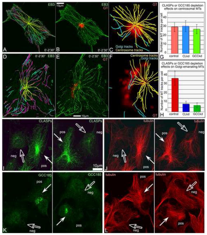
A, Western blotting illustrating ∼75% decrease of both CLASP1 and CLASP2 on the 3rd day after siRNA transfection as well as expression of non-silenceable GFP-CLASP2. Regions of interest are shown. B. Numbers of non-centrosomal MTs formed 45″ after nocodazole washout in CLASP-positive (red, 30 cells) and CLASP-depleted cells (blue, 30 cells), as well as upon rescue by non-silenceable GFP-CLASP2 (green, 16 cells). Based on tubulin immunostaining. C-F, Mixed culture of CLASP-positive (pos) and CLASP-depleted (neg) cells 45″ after nocodazole washout. Tubulin (red), GM130 (blue), CLASPs (green). Immunostaining. C, In CLASP positive cell, MTs radiate from Golgi stacks (arrows). D, In CLASP-depleted cell, Golgi stacks are not associated with rare MTs (hollow arrows). E, CLASPs and F, MTs and Golgi stacks at low magnification. Boxes enlarged in C and D. G, H. Video frames illustrating MT formation at the Golgi (arrows) in mRFP-EB3 (false-colored green) and YFP-GT (false-colored red) in nocodazole washout. Time after nocodazole removal in shown. Left, frames showing whole cell. Areas in boxes are enlarged to the right. G, Control cell. H, CLASP-depleted cell.