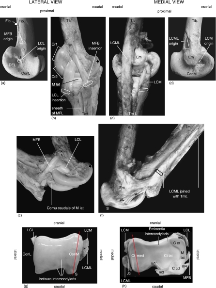
Fig. 3.
Anatomical photo reference in accordance with Table 2, left limb. (a) Macerated distal tibiotarsus, lateral view. (b) Intertarsal joint at maximum extension, lateral view. Black dot on trochlea cartilaginis tibialis indicates reference point. (c) Intertarsal joint in flexion. Black dot indicates reference point (see b). (d) Macerated distal tibiotarus, medial view. (e) Intertarsal joint at maximum extension, medial view. (f) Intertarsal joint in flexion with osseous protrusion (S) to prevent hyperextension and tensed broad ligament (BL) preventing LCML from sliding over Em. (g) Tibiotarsal joint surface with LCL, LCM and LCML severed. (h) Tarsometatarsal joint surface with LCL, MFB, LCM and LCML severed. Joint capsule (Jc) associated with LCML.

