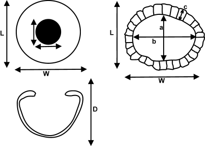
Fig. 2.
Schematic of the surface view (top left) and lateral view (bottom left) of a modern shark scleral skeletal cup, and the surface view of a fossil scleral skeletal ring (top right). L, length of the scleral cup; W, width of the scleral cup; D, depth of the scleral cup; a, length of the aperture; b, width of the aperture; c, width of scleral skeletal plate/ring.

