Figure 2.

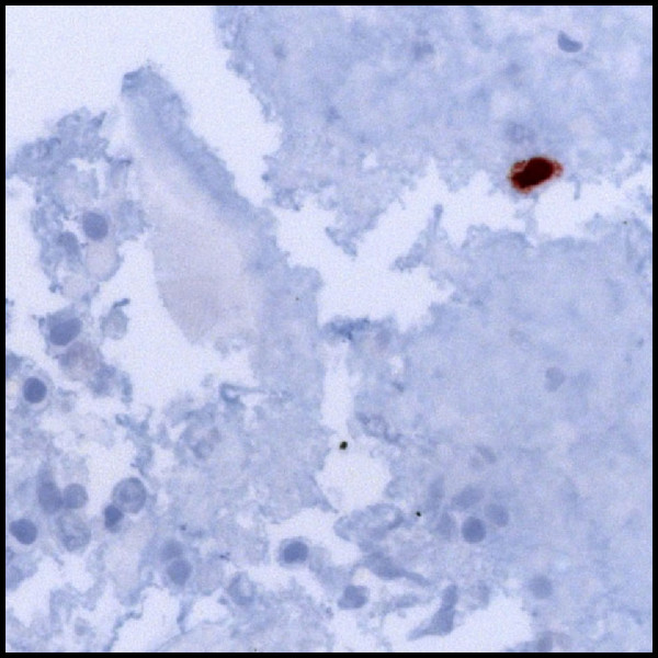
Immunohistochemical staining for HSV-1 in a fixated cyotspun obtained from BAL from Case 2 identifying a positive cell (dark brown colour). Bottom and left: alveolar macrophages and lymphocytes with degenerative changes.

Immunohistochemical staining for HSV-1 in a fixated cyotspun obtained from BAL from Case 2 identifying a positive cell (dark brown colour). Bottom and left: alveolar macrophages and lymphocytes with degenerative changes.