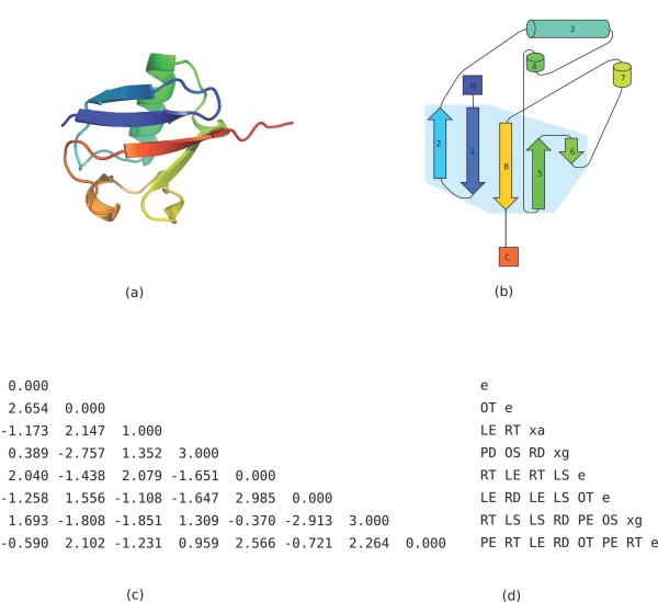
Figure 13.
β-grasp query structure and tableau. (a) 3D structure of ubiquitin, PDB identifier 1UBI. Image generated with PyMOL. (b) Topology cartoon of 1UBI. (c) Orientation matrix Ω for 1UBI. Each angle is in radians between -π and π. The main diagonal denotes the SSE type by 0.000, 1.000, 2.000, or 3.000 for β-strands, α-helices, π-helices, and 310-helices, respectively. (d) Tableau for 1UBI. The main diagonal denotes the SSE type by e, xa, xi, or xg for β-strands, α-helices, π-helices, and 310-helices, respectively.

