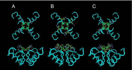
Fig. 2.
Molecular dynamics. Top and side wire frame views of calix[4]arenes 1 (A), 4 (B), and 8 (C) docked into the X-ray structure of the human voltage dependent Kv1.2 potassium channel. Only the backbone helices of the α-subunits are shown. The Asp-379 side chains are represented, showing the 4-ion-pair, hydrogen-bonded contacts with the guanidinium residues of the calix[4]arene. Structures were optimized in vacuo at 300 K applying positional restraints to Kv1.2.

