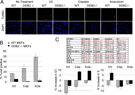
Fig. 1.
DDB2−/− MEFs exhibit cell cycle arrest and apoptosis defect upon DNA damage. (A) Wild-type or DDB2−/− MEFs were treated with UV-C (50 J/m2) or cisplatin (30 μm) or aclarubicin (0.5 μM) for 24 hours. After the treatments, the cells were subjected to TUNEL assay using the ApopTag Red In Situ Apoptosis Detection Kit and procedure provided by the manufacturer (Chemicon International). Average percentages of the TUNEL-positive nuclei from 10 different fields from two independent experiments are plotted (B). (C) After treatment with the DNA-damaging drugs, cells were subjected to flow-cytometric analyses. An average of the cell cycle distribution (including the SubG1 cells) from three different sets is shown.

