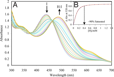
Fig. 2.
Example of UV/vis titration. (A) UV-vis titration of host (S)-6 with PV (150 μM). (B) The 1:1 binding isotherm (plot of the difference in absorbance at 520 nm with the addition of the host). [H]t, total host concentration. All titrations were carried out in 100% MeOH, 10 mM para-toluenesulfonic acid and Hunig's base buffer (pH 7.4). All measurements were taken at 25 °C. The solid line is the calculated curve resulting from iterative data fitting to a 1:1 binding isotherm (12).

