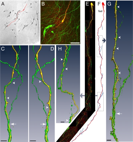
Fig. 1.
Biocytin-filled GnRH neurons exhibiting dendro-dendritic bundling with other GnRH neurons in the verticle orientation. (A) Montage of low power confocal images through a thick slice of the rPOA shows GnRH-GFP neurons (black) and a single GnRH neuron that was filled with biocytin and subsequently labeled with a fluorescent marker (traced in red). (B) High power projection of confocal images of the filled neuron in A, showing endogenous GFP expression in multiple GnRH soma and in a plexus of processes (green) and fluorescently labeled, biocytin-filled GnRH neuron (yellow/red). (C and D) Three-dimensional isosurface rendering of GFP (green) and biocytin-filled (yellow) dendrites (rectangle in A) that bundle together viewed from 2 angles 180° apart. Arrowhead indicates 2 bundled dendrites; arrow shows the bundling of 3 dendrites. (E) Montage of high power confocal image stacks showing another filled GnRH neuron exhibiting dendritic contacts and bundling in the vertical orientation. (F) A schematic traced from confocal image data illustrates 15 apparent close associations between the filled dendrite (red) and other GnRH neuron dendrites (blue). Three-dimensional rendered images of the filled dendrite and apposing dendrites created from high power confocal images are shown for the highlighted regions. (G) Two juxtaposed GnRH neuron dendrites can be seen in the upper portion of the image (arrowheads), and 3 dendrites are found bundling together further down the length of the dendrite (arrow). (H) Points of contact are apparent between the filled dendrite and 2 dendrites that run in parallel with the filled dendrite (arrowheads). (Scale bars: B, 50 μm; C, D, G, and H, 5 μm.)

