Fig. 2.
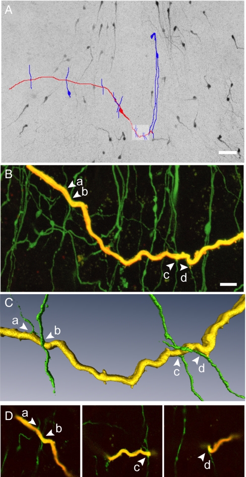
Dendro-dendritic associations in the horizontal orientation. (A) Montage of low power confocal images through a thick brain slice through the rPOA shows GnRH-GFP neurons (black) and a single GnRH neuron that was filled with biocytin and subsequently labeled with a fluorescent marker (traced in red). GnRH-GFP neuron dendrites in apparent close apposition with the dendrite of the filled GnRH neuron are traced in blue. (B) High power confocal projection of the area highlighted in panel A showing a portion of the filled dendrite (yellow) and other endogenously fluorescent GnRH processes. Arrows indicate 4 dendrites (a–d) found in close apposition with the filled dendrite. (C) Isosurface rendering of the dendrites in close apposition show the perpendicular nature of these interactions. (D) Individual optical sections (0.32 μm thick) showing the close apposition of dendritic processes. (Scale bars: A, 50 μm; B, 10 μm.)

