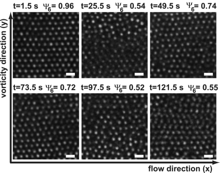
Fig. 2.
Confocal microscopy images taken in the velocity–vorticity plane of an initially crystalline suspension of 1.2-μm diameter silica particles in ETPTA sheared with γ̇ = 2 s−1. Initially, the structure was crystalline, but it became largely disordered, although ordered domains kept forming temporarily. (Scale bars, 2 μm.)

