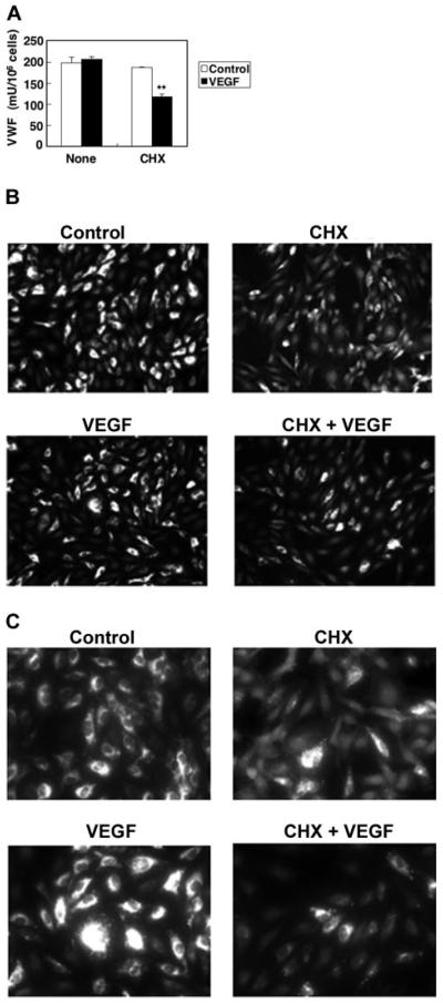
Figure 2. Kinetics of VWF expression in VEGF treated cells.
(A) Steady-state VEGF protein levels in HAECs. HAECs were treated with 50 ng/mL VEGF or 10 μM CHX 10 μM for 2 hours. The concentration of VWF in the cells was measured by an ELISA. (n = 3 ± SD; *P < .01 compared with VEGF alone). (B) Immunofluorescence of VWF in endothelial cells, low magnification. HAECs were treated with 50 ng/mL VEGF or 10 μM CHX for 2 hours and then stained with anti-VWF antibody and Cy3-conjugated IgG secondary antibody. Magnitude, × 100. (C) Immunofluorescence of VWF in endothelial cells, high magnification. HAECs were treated with 50 ng/mL VEGF or 10 μM CHX for 2 hours and then stained with anti-VWF antibody and Cy3-conjugated IgG secondary antibody. Magnification, × 400. Cells were imaged at 22°C under a Nikon Fluorescent Eclipse E600 microscope (Nikon, Tokyo, Japan) equipped with 10 × 10 and 10 × 40 water immersion objective lenses (Nikon). Images were acquired with a Nikon DXM 1200 digital camera and analyzed with Spot Epi-fl 3.5.2 software (Diagnostic Instruments, Sterling Heights, MI).

