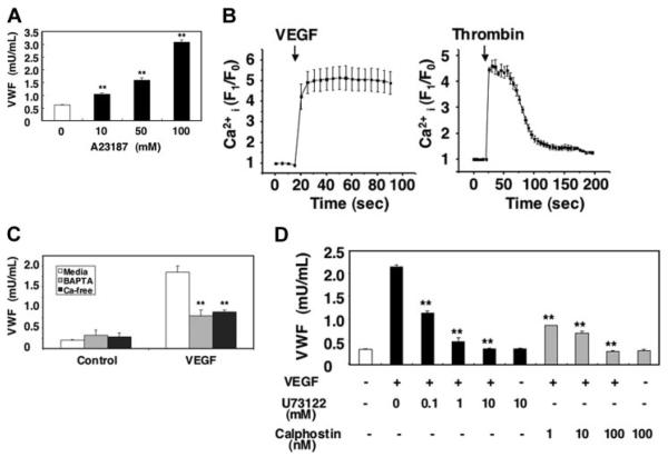
Figure 3. Long-term treatment with VEGF inhibits VEGF-induced exocytosis.
(A) The calcium ionophore A23187 activates VWF release from endothelial cells. HAECs were incubated with A23187 for 1 hour. The amount of VWF released from cells into the media was measured by an ELISA (n = 3 ± SD; **P < .01 versus 0 μM). (B) VEGF increases intracellular calcium levels. HAECs were incubated with the calcium indicator fluo-3AM for 20 minutes. The integrated fluo-3 fluorescence intensity of each cell was measured in real time after treatment with 50 ng/mL VEGF or 1 U/mL thrombin (n = 3 ± SD). (C) Calcium pools and VWF release from endothelial cells. HAECs were pretreated with 20 μM BAPTA/am for 30 minutes in DMEM or Ca2+-free DMEM and then incubated with 50 ng/mL VEGF for 1 hour. The amount of VWF released from cells into the media was measured by an ELISA (n = 3 ± SD; **P < .01 versus media + VEGF). (D) PLC and PKC inhibitors block VEGF-induced VWF release from endothelial cells. HAECs were pretreated with the PLC-γ inhibitor U73122 for 30 minutes or the PKC inhibitor calphostin C for 30 minutes and then incubated with 50 ng/mL VEGF for 1 hour. The amount of VWF released from cells into the media was measured by an ELISA (n = 3 ± SD; **P < .01 versus no inhibitor).

