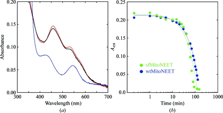
Figure 1.
UV–visible spectra of the 2Fe–2S cluster of mitoNEET. (a) Absorption spectra of mitoNEET (33–108) purified from fusion protein recorded from 300 to 700 nm (red spectrum). Upon reduction by the addition of sodium dithionite (blue spectrum), the typical A 458 peak is greatly reduced. Re-oxidation of the 2Fe–2S cluster occurs upon exposure to oxygen and is indicated by restoration of the typical A 458 peak (black spectrum). This is identical to the behavior of the 2Fe–2S cluster in mitoNEET described previously (Paddock et al., 2007 ▶; Wiley, Paddock et al., 2007 ▶). (b) Time-dependent decay of the 2Fe–2S cluster at pH 6. MitoNEET purified from the sfGFP fusion (sfMitoNEET; green) is overlaid with wild-type mitoNEET purified without fusion (wtMitoNEET; blue). Results are consistent with those previously reported (Paddock et al., 2007 ▶; Wiley, Paddock et al., 2007 ▶).

