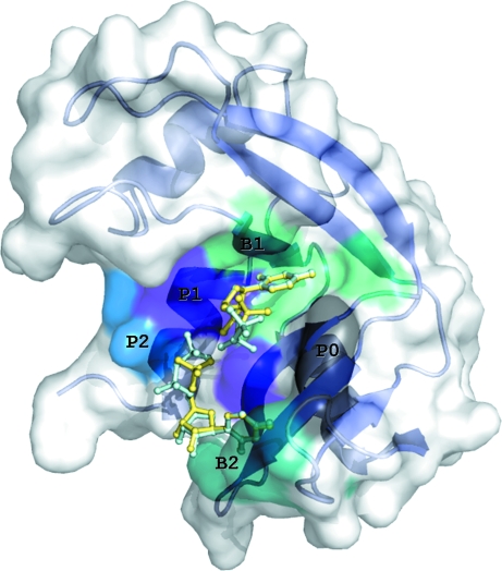
Figure 1.
A schematic diagram of the RNase A molecule with U5P (yellow) and UDP (cyan) molecules superimposed bound at the active site. The molecular surface and secondary structure of the enzyme are also shown. Subsites P0 (Lys66), B2 (Asn67, Gln69, Asn71, Glu111, His119), P1 (Gln11, His12, Lys41, His119), B1 (Val43, Asn44, Thr45, Phe120, Ser123) and P2 (Lys7, Arg10) are labelled and marked on the molecular surface with different colours.

