Fig. 3.
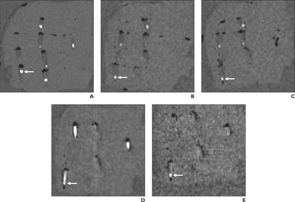
CT scans of phantom with multiplanar reconstruction parallel to long axis of cone-shaped plastic container in right lower quadrant of phantom containing calcium hydroxyapatite stone (arrows) that is 3.8 mm in short-axis diameter. Constant scanning parameters were 64 × 0.6 mm collimation with 1-mm reconstruction slice thickness and without 4-cm-thick oil gel surrounding phantom. Stone in plastic container in right lower quadrant (arrows) is detectable on all scans except scan obtained with 80-140 kVp pair and 107 mg/dL iodine solution combination (D). Note severe degree of residual nonsubtracted iodine with this combination obscures stone. Scan with 100-140 kVp pair and 107 mg/dL iodine solution combination (E) shows mild degree of residual nonsubtracted iodine, but stone is visible. Note stone appears smaller on dual-energy CT scans with virtual unenhanced iodine subtraction (B, C, and E) than on true unenhanced scan (A). Due to slight difference in orientation of phantom between scans, other stones and plastic containers are variably seen.
A, Single-energy CT scan (120 kVp) with plastic container filled with saline (true unenhanced scan).
B-D, Dual-energy CT scans (80-140 kVp pair) with plastic container filled with 21 (B), 64 (C), and 107 (D) mg/dL iodine solution, respectively, with virtual unenhanced iodine subtraction.
E, Dual-energy CT scan (100-140 kVp pair) with plastic container filled with 107 mg/dL iodine solution with virtual unenhanced iodine subtraction.

