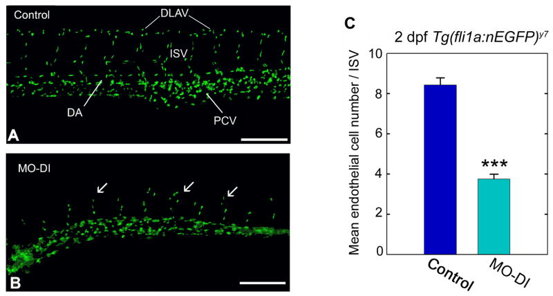
Fig. 1.
Perlecan knockdown influences endothelial cell number. Vascular analysis of control (A) or perlecan morphant (B) Tg(fli1a:nEGFP)y7 zebrafish embryos. Notice the abnormal formation of ISVs (arrows) and the lack of a DLAV in the perlecan morphants. (C) Endothelial cell number was assessed by counting the endothelial cell nuclei present per intersegmental vessel region in 2 dpf control and morphant embryos. Control embryos typically displayed eight endothelial cells/ISV region compared to perlecan morphants which typically displayed four cells in the comparable area. Data summarize the endothelial cell counts from a total of 40 ISV regions for controls and a comparable 40 ISV regions for morphants. The forty counts were derived from four separate embryos, with 10 ISV regions assessed in each (***P<0.001). Scale bar, 250 μm.

