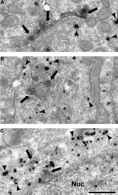
Figure 2.
Electron micrographs of cell bodies immunogold labeled for PV or CR (black arrowheads) which also contain DAB label (black arrows) for D1 and D5. In PV somata, the stereotypical D1 staining of the Golgi apparatus was identified (A), as well as labeling associated with other internal membrane structures, including endoplasmic reticulum and mitochondria (B). In CR somata, D5 staining was associated with internal membranes (C). Nucleus (Nuc). Scale bar is 500 nm.

