Figure 3.
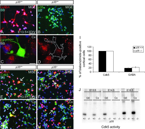
Cdk5 is expressed, but not active, in p35-deficient interneurons. Cultured neurons from E13.5 MGE (A, B, E, F) or Cx (C, D, G, H), immunoreacted for GABA (A), p35 (B–D), or GABA/Cdk5 (E–H), and stained with bisbenzimide (BB) to show nuclei. The majority of p35+/+ MGE cells express GABA (A) and p35 (B). p35 Is also present in GFP+ cortical interneurons obtained from GAD67GFP/+ mice (C, D). Cdk5 is expressed in GABAergic MGE (E, F) and Cx (G, H) neurons (arrows) regardless of genotype. (I) Graph shows the proportion of cortical cells expressing Cdk5+ and GABA+ in dissociated cell cultures. No significant difference in GABA expression is observed between control and p35−/− mice. (J) Cdk5 was isolated from cell lysates of GE and Cx by immunoprecipitation and its activity measured by an in vitro kinase assay in the presence of histone H1. Cdk5 activity is undetectable in embryonic GE and Cx of p35−/− compared with p35+/− mice. M, medial; Cx, cortex; DIV, day in vitro. Error bars represent SEM. Scale bar, 50 μm (A, B, E–H), 10 μm (C, D).

