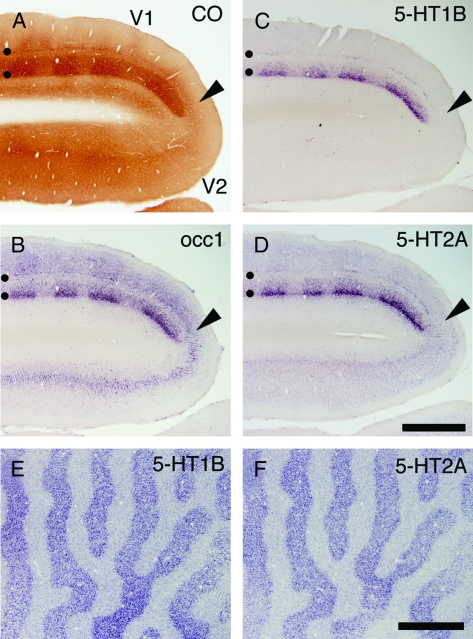
Figure 6.
Expressions of 5-HT1B and 5-HT2A receptor mRNAs in V1 following monocular inactivation. TTX was injected into 1 eye twice a week for 21 days (A–D), or 3 h before sacrifice (E,F). These monocularly inactivated monkey brains were histochemically analyzed. (A) CO staining near V1-V2 border (shown by the black arrowheads). (B–D) ISH in V1 and V2. (B) occ1, (C) 5-HT1B receptor, (D) 5-HT2A receptor. Adjacent sections were analyzed in this order. Black circles in panels (A–D) indicate layers IVA and IVCbeta. (E and F) 5-HT1B and 5-HT2A receptor mRNA expressions were examined by ISH after 3 h monocular inactivation (MD). These photos show tangential sections at the level of layer IVC. Note that 5-HT1B and 5-HT2A receptor mRNAs exhibit almost identical patterns. Scale bars: 500 μm.

