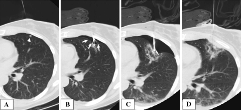
Fig. 1.
Sequential CT images of radiofrequency ablation of subpleural tumor. A A 22-gauge Chiba needle was placed adjacent to the peripherally located metastasis (white arrowhead: subpleural tumor). B An intentional and iatrogenic artificial pneumothorax was created displacing the visceral pleura (white arrows: artificial pneumothorax). C Enlargement of the artificially created pneumothorax further separated the ablation site from the parietal pleura. D Following completion of the ablation, the tines were retracted and a localized pneumothorax was decompressed by active aspiration using a syringe

