
Figure 4.
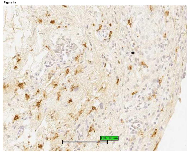
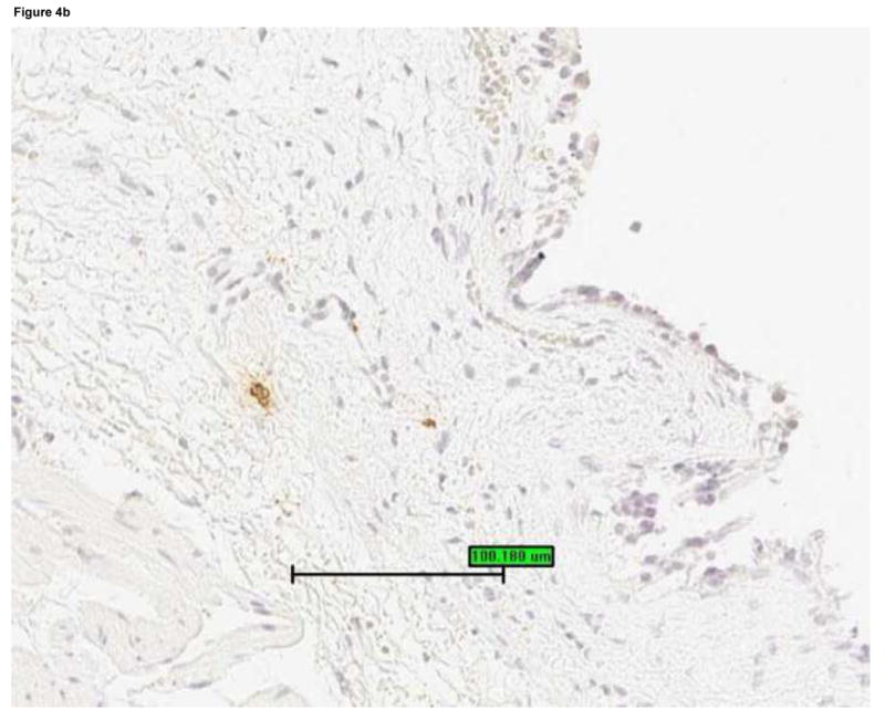
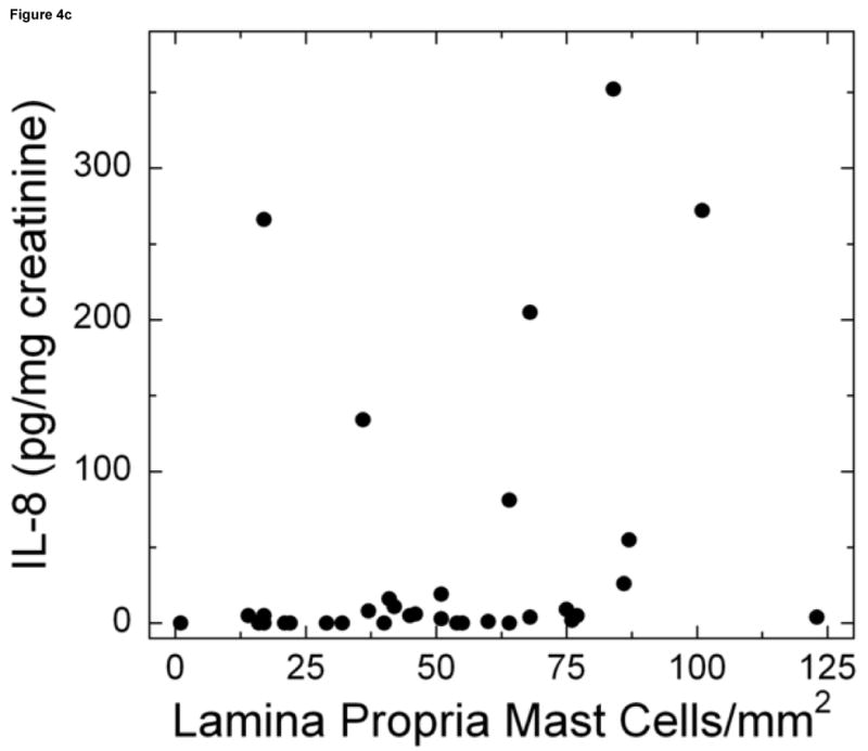
Mast cells were detected by staining bladder biopsies with anti-tryptase primary antibody followed by a standard streptavidin method. An optical reticle was used to count mast cells in the urothelium, lamina propria and detrusor. a: Brisk mastocytosis in the lamina propria (magnification 200×). b: Minimal mastocytosis in the lamina propria (magnification 200×). c: Scatter plot of urine IL-8 levels vs. mast cell count in the lamina propria for untreated IC/PBS patients. Lamina propria mast cell count was significantly associated with urine IL-8, which was analyzed by ELISA and normalized to urine creatinine.