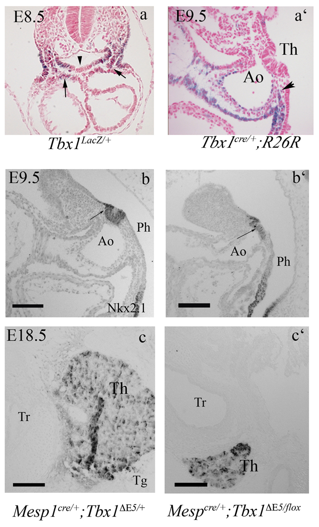
Figure 3. Thyroid development requires Tbx1 expression in the mesoderm.
(a) Transverse section of an X-gal stained, Tbx1lacZ/+ E8.5 embryo showing Tbx1 expression in the mesenchyme adjacent to the thyroid primordium (arrows) but not in the primordium itself (arrowhead). (a’) cell fate analysis in Tbx1Cre/+ ; R26R mice. A group of β-gal+ cells surrounds the thyroid primordium (black arrow) but no labeling is visible within the primordium. (b–c) The thyroid phenotype in Mesp1cre/+;Tbx1flox/− embryos at E9.5 and E18.5. Nkx2-1immunohistochemistry on sagittal sections of mutant embryos at E9.5 (b’) reveals a differentiated thyroid primordium that is smaller and flatter than in controls (b). (c–c’) Tg immunohistochemistry of Mesp1Cre/+ ;Tbx1flox/− mutant embryos at E18.5. Ao aortic arch, Tr trachea. The scale bar is 100µm.

