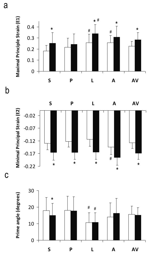
Figure 4.
Maximal (a) and minimal (b) principal strains and the prime angle between the primary eigenvector and the radial direction (c). S, Septum; P, posterior; L, lateral; A, anterior; AV, slice average. White and black bars are sub-epicardium and sub-endocardium, respectively. *P<0.05 sub-epicardium versus sub-endocardium. #P<0.05 comparing with other segments.

