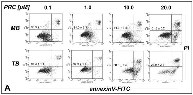
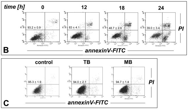
Figure 2. Preferential induction of apoptosis by PRC compounds in malignant G361 melanoma cells.
(A) Dose-response relationship (24 h continuous exposure) of induction of G361 melanoma cell apoptosis by the PRC compounds MB and TB (0.1–20 μM, each) were established by flow cytometric analysis of annexinV-FITC/propidium iodide-stained cells. (B) Time course of TB-induction (10 μM) of G361 melanoma cell apoptosis. (C) No induction of cell death was observed when normal human skin fibroblasts (Hs27) were exposed to MB (20 μM) or TB (10 μM, 24 h continuous exposure). Early apoptotic and late apoptotic/necrotic cells are located in the lower right (AV+, PI−) and upper right quadrant (AV+, PI+), respectively. One representative experiment of three similar repeats is shown. The numbers indicate viable cells (AV−, PI−, lower left quadrant) in percent of total gated cells (mean ± SD, n=3).


