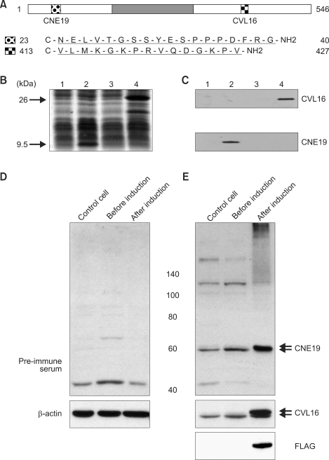
Figure 2.
ALADIN-specific peptide antibodies. (A) Antigen sequences of ALADIN. The dark grey box represents the WD repeat domain. (B) Protein extracts from Escherichia coli cells expressing a His-tagged N-terminal 33 aa ALADIN sequence (lanes 1 and 2) and a His-tagged C-terminal 166 aa ALADIN sequence (lanes 3 and 4) were separated on 15% (w/v) SDS-PAGE. Lanes 1, 3: lysates prior to induction; 2, 4: lysates after induction. Proteins were stained with Coomassie Brilliant Blue. Arrows indicate the induced His-tagged proteins. (C) The CNE19 antibody recognized a 9.6 kDa protein, while the CVL16 antibody recognized a 26 kDa protein. (D) and (E) Western blot analysis of ALADIN in HeLa cells. FLAG-tagged ALADIN was expressed using Tet-off system as described previously (Min et al., 2003). Upper and lower arrows indicate FLAG-ALADIN and endogenous ALADIN, respectively.

