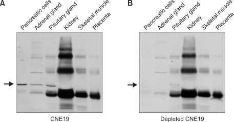
Figure 3.
Expression of ALADIN in human tissues. Fifteen µg of protein from tissues and primary cells lysates were separated on SDS-PAGE. (A) Western blot analysis using anti-CNE19 antibody. The arrow indicates the expression of ALADIN in pancreatic cells, adrenal and pituitary gland cells. (B) Western blot analysis using adsorbed CNE19 antibody. The arrow shows where ALADIN bands (now absent) would be expected.

