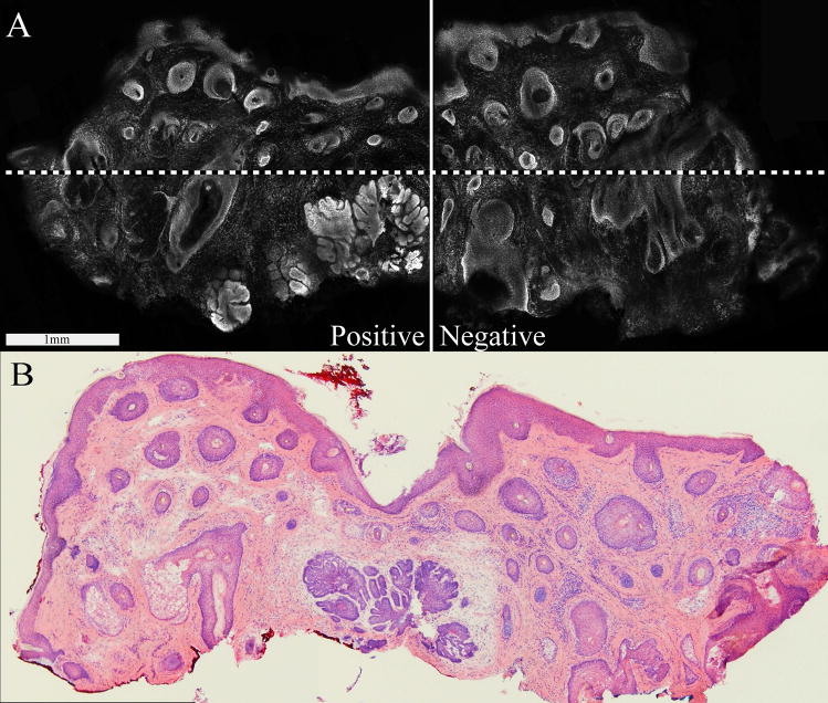
Figure 2.
Examples of morphological features as seen in confocal fluorescence submosaics and the corresponding Mohs frozen histopathology. In each pair of images, the left one is the confocal and the right is the histopathology. Benign features are: (A–B) eccrine gland, (C–D) sebaceous gland, (E–F) epidermis (epi) along the periphery of the Mohs excisions containing hair follicles (h), along with lymphocytic inflammatory infiltrate (ii) in the deeper surrounding dermis. Malignant features are: (G–H) nodular BCC tumor, (I–J) micronodular BCC tumor and (K–L) infiltrative BCC tumor.

