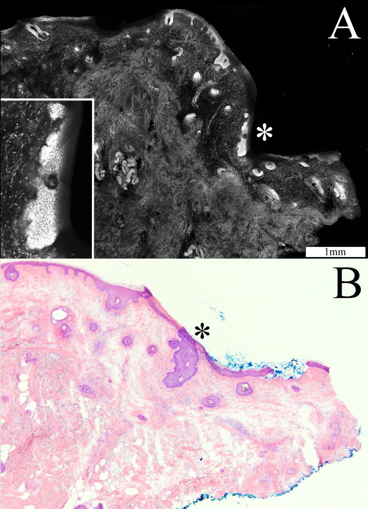
Figure 3.
Small infiltrative BCCs are differentiated from normal features such as hair follicles in the confocal mosaic (A), as validated by the corresponding histopathology (B). Enclosed in the dotted circle is the cluster of infiltrative tumors. The large cluster in this case made the tumors easily identifiable.

