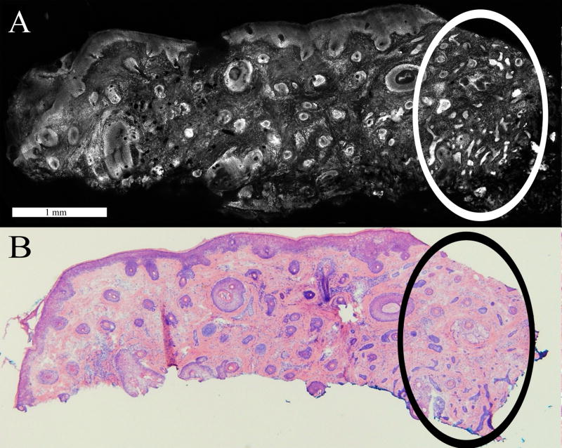
Figure 4.
Superficial BCC tumor (*) extending contiguously from the epidermis along the periphery of the Mohs excision, as seen in the confocal mosaic (A) and corresponding histopathology (B). The inset, at high magnification, shows nuclear crowding, peripheral palisading and leaf-like projections from the epidermis.

