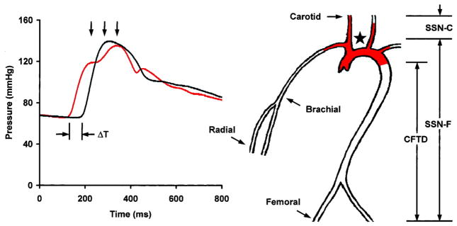
Figure 1.
Measurement of PWV. Transit time, ΔT, between the foot of the carotid (red waveform) and femoral waveforms is measured with a tonometer. Carotid-femoral transit distance (CFTD) is estimated by measuring the distance from the suprasternal notch (SSN, ) to the carotid (SSN-C) and femoral (SSN-F) sites and taking the difference to account for parallel transmission along the brachiocephalic and carotid arteries and around the aortic arch (red shading). This corrected distance is divided by transit time delay to give PWV. Note that carotid-femoral PWV fails to assess stiffness of the proximal aorta (red shading). (Reproduced from (62).)

