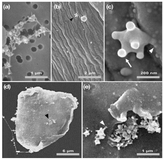
Fig. 2.
Scanning electron micrographs of L. heterotoma VLPs. (a) In untreated fluid, VLPs remain connected to one another. (b, c) VLPs on the surface of a wasp egg, 10–15 min after wasp infection. (d, e) Binding of VLPs to a lamellocyte after 30 min incubation. The arrowhead in (d) shows a cluster of VLPs; this area is enlarged in (e). The white arrowheads in (c) and (e) show the spike/knob structure of a VLP in contact with the surface of a wasp egg (c) and the surface of a lamellocyte (e), respectively. The arrowheads in (b) and (d) point to areas enlarged in (c) and (e), respectively.

