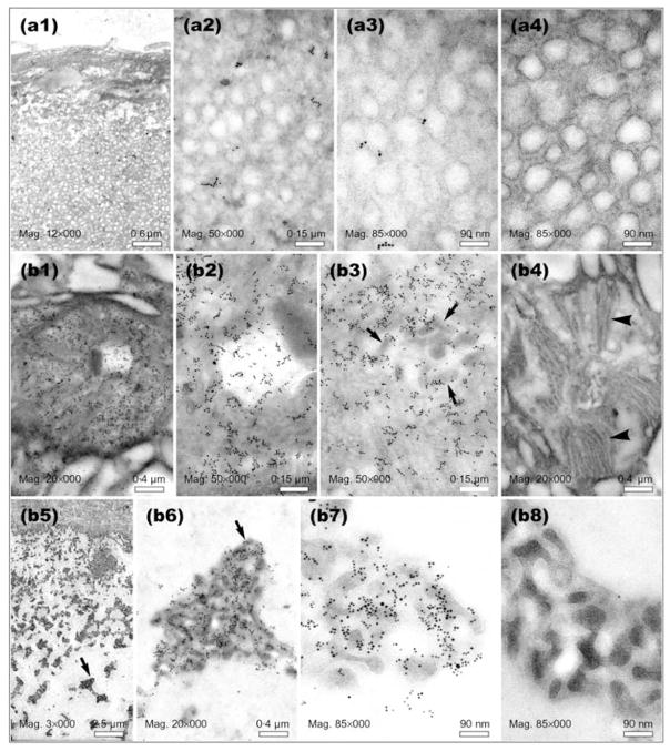
Fig. 4.
Immuno-EM of thin sections prepared from positions A and B of the L. heterotoma long gland (see Fig. 1d for orientation). Panels a1–a4 show preparations derived from the ‘nose’, panels b1–b4 through the ‘rough’ canals within secretory cells and b5–b8 through the lumen of the long gland. Panels a4, b4 and b8 are negative controls (no primary antibody). Arrows in panels b3, b5 and b6 point to clusters of immature VLPs. Arrowheads in panel b4 point to membranous projections found around the ‘rough’ canals into which immature VLPs are first secreted. Similar structures were found in secretory cells in the long gland at position C (see Fig. 5, panel c3, black arrowhead).

